I didn’t expect this blog to bleed into my optic life. But, this is exactly what happened last week!
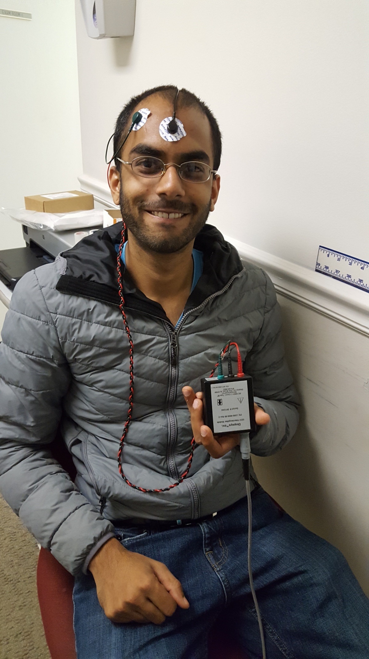
For the first time in my life, I had electrodes attached to my forehead for brain-imaging. The tech this time was VEP, short for “visual evoked potential”. (More specifically, what I sat for was the Diopsys® NOVA-VEP test.)
I was in my local optometrist’s clinic for a couple of eye exams. I’d lost my glasses, and had nearly run out of contacts: so, I needed replacements all-around.
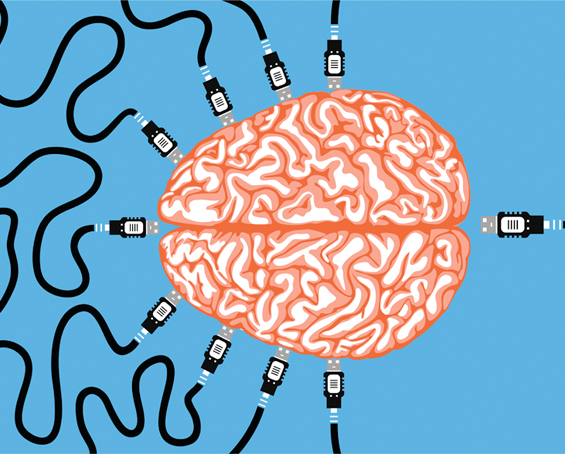
VEP measures electrical activity taking place in the brain’s occipital lobe. Breaking VEP down, we have:
- “Visual” — patients observe provided visual stimuli;
- “evoked” — electrical energy is generated at the retina;
- “potential” — electrical activity is measured in the visual cortex
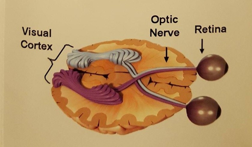
Simplified image of the two main neural-optic pathways
I will leave this post shorter than you can expect others in this series to be. My encounter with VEP was brief and unanticipated, but the main fun was having the electrodes attached to my forehead.
Now, if only I had my VEP brain scan on-hand…
Sources:
- Diopsys NOVA-VEP® and Diopsys NOVA-ERG®
- Visual Evoked Potential brochure (procured from Dr. Tillman’s clinic in Carrollton, Georgia, U.S.A.)